TRD 4: Discovery Systems: Visualization, Modeling, and Workflow
The Resource has a mix of applied computer science and biomedical microscopy applications, producing specialized visualization and analysis tools targeted primarily at optical and scanned-probe microscopy systems. Our collaborators continue to provide challenging visualization and analysis problems that drive technique development and dissertations in computer science. Our focus on solving collaborators’ problems by providing computer-integrated tools continues to result in new visualization and analysis tools.
The difficulty with toolkits like the open-source Visualization ToolKit (VTK) from a scientist’s point of view is rather like the difficulty a computer scientist would have if given free access to an automotive shop: all the tools needed to fix a car are in there somewhere, but knowing which tools to use and how is beyond their experience. Our aim is to provide tools that are optimized to answer the specific questions posed by our collaborators in the belief that a tool that solves a particular problem is more likely to be of broader usefulness than a “generic” tool that is not optimized for usefulness on any particular problem. This requires drawing on an understanding of the available techniques, the characteristics of the human visual system, the characteristics of the data sets, and the questions being asked.
Our collaborators continue to provide data sets and questions that push the state of the art in 3D visualization. Problems generated from Radiology colleagues have produced two computer-science dissertation topics and tools of use for both micron-scale and organ-scale data sets. We have four ongoing projects in this area, with the longest-running one (ImageSurfer) maturing into a visualization and analysis tool and the two newest ones (nDive and FOR) providing publications and novel views into biomedical and microscopy data sets. We continue to apply cutting-edge computer graphics and interaction to biomedical problems in the Eve for Microscopy project.
3D Interactive Protein Structure Formation: SketchBio.
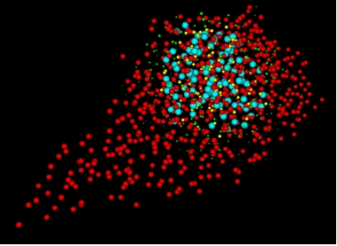
Multivariate volume data with uncertainty: nDive.
3D Scalar Volume Visualization: ImageSurfer
Advanced Graphics and Interaction, including EVE for Microscopy