Project 2.2
Project 2.2. Develop broadly applicable approaches to confer light-induced activation and release of proteins in living cells, and use these to generate localized changes in protein activity at precise positions and times relative to applied forces. Published methods to control the activity of GTPase proteins in the Rho family with light are used to alter the kinetics and subcellular location of GTPase activities during the application of forces to living cells. We also develop new methods that make it possible to easily generate photo-regulated analogs of many proteins of interest. These include LOVTRAP, reversible light-induced release of proteins sequestered at internal membranes, and the caging of bio-active peptides that control the activity of endogenous target proteins.
http://www.hahnlab.com/tools/controlProt-Light.html
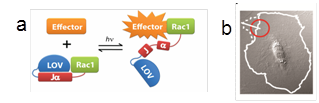 Fig. 6. Photoactivatable Rac (PA-Rac). a) Cartoon representation of Rac caging by the LOV
domain. When in the dark, the Jα portion of LOV is an alpha helix. Upon irradiation it unwinds to release steric inhibition of Rac. b) Pulses of irradiation restricted to the red circle produce localized Rac activation and therefore localized protrusion (cell outline changes from solid to dotted line).
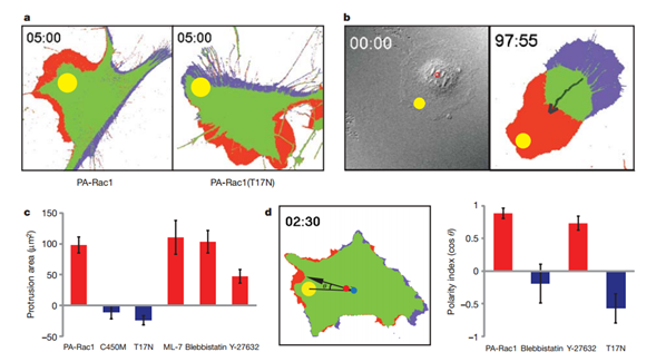
Figure 2 | Localized activation or inactivation of PA-Rac1 induces myosindependent migration. a, Protrusion/retraction map after a single pulse of activating illumination. MEFs expressing PA-Rac1 (left) generated protrusions at the site of irradiation (red) and retraction at the opposite side of the cell (blue) (in all 50 cells studied). Irradiation of the dominantnegative T17N mutant of PA-Rac1 (right) produced retraction near the point of irradiation, with protrusion in area(s) other than the site of irradiation (in all 25 cells studied). b, Repeated activation of PA-Rac1 at the cell edge induces directional migration. (MEF, 2-min intervals, average 0.8 mm movement per pulse, n5 6.) c, Localized activation of PA-Rac1 in the presence of ML-7 (MLCK inhibitor, 1mM), blebbistatin (myosin II ATPase inhibitor, 1mM) or Y-27632 (ROCK inhibitor, 10mM). Protrusions analysed as in panela. d, Effectof myosinor ROCKinhibition on the ability of Rac1to specify the direction of movement. The cosine of the angle between two lines (from the irradiation spot to the cell centroid at time 0, from the centroid at time 0 to the centroid at the end of the experiment) indicated how much the cell deviatesfrom thedirection specifiedby localirradiation.For c,d, n. 25; means6 95% confidence intervals; throughout Fig. 3 irradiation at 458nm, spot diameter5 10mm; time shown is in minutes and seconds.
Nature, 461: 104-110, 2009. PMC2766670
Meth. Enzym., 497: 393-407, 2011. PMC3407667
examples of applications in animals include: Nat Cell Biol., 12: 591-7, 2011. PMC2929827; Dev Cell. 18: 226-36, 2010. PMC2824622; Nat Neurosci. 15: 891-6, 2012. PMC3565539