Project 1: High Throughput Technologies
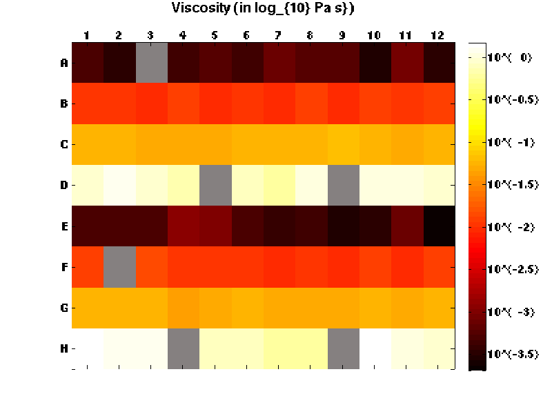
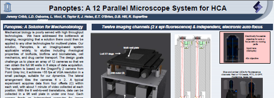
 High Throughput Microscopy: Panoptes is a unique and custom-designed high-throughput array microscopy system capable of acquiring data on twelve-channels simultaneously all while using SBS standard 96-well or 384-well plates. Each channel in the system can independently drive two-channel fluorescence imaging at up to 51 frames per second. We use Panoptes to study mucus rheology, clot permeability, and cell mechanics—it can perform these experiments, with completed data analysis, on 96 specimens in approximately six hours, with no user attention. View a fly-through of the system design or download our poster.
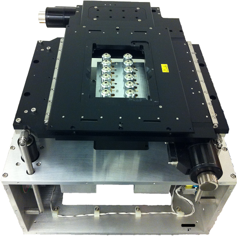
The entire system, designed by CISMM engineer Leandra Vicci, comprises 77 custom mechanical parts and 21 custom electronic boards. Fifteen of the electronics boards control signal synchronization and are housed in an electronics box (not shown). The remaining custom boards, located within the Panoptes base, provide camera control, sample illumination, and z-alignment/positioning. Twelve 40x objectives, located underneath twelve wells of the multiwall plate, add fine, motionless control of the imaging plane through the use of liquid lenses. A Nanometric stage moves the multiwall plate in X and Y, visiting all of its 96-wells in just seven steps. Three stepper motors support the Nanometric stage and provide fine approach and tilt adjustment that makes the plane of the multiwell plate parallel to the plane of the objectives. This adjustment is needed for each sample plate used in the system due to the uncontrolled plate distortion which can be as large as several hundred microns.
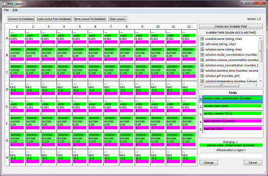
 The experiment begins with a well layout program that allows the user to input the metadata for each well, and to specify the parameters for executing the experiment including, for example, the camera and illumination specifications, the number of fields of view to visit per well, and the well-to-well trajectory through the plate. The experiment then proceeds with Panoptes image acquisition. Typical parameters use two-color epi-fluorescence video capture at 55 fps, 1 minute of video in each of five fields of view per well, with seven larger plate translations to acquire data over all 96 wells. This automated data acquisition step typically requires less than one hour.

When finished with data collection, the system analyzes the acquired video, starting with CISMM’s Video Spot Tracker software for most experiments. The Video Spot Tracker application identifies and tracks particles that satisfy the signal threshold, obtaining time series of positions for the beads in the fields of view. Doing this for the nearly 1 Tb of video requires approximately 5 hours to complete for a typical experiment providing trajectories for about 5000 beads.
Finally, the particle trajectories provided by Video Spot Tracker are processed by the subsequent statistical analysis, programmed in Matlab. All steps, from acquisition through analysis, take a total of about 6 hours to complete. Panoptes automates this entire process through computer control, making it a fully autonomous high-throughput microscopy system.
