TRD 1: Force Microscope Technologies
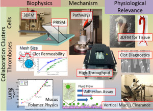
Mechanobiology can be broadly defined as the study of the role of forces, force response, and mechanical properties in biological systems. Within the cell, forces transport vesicles, dictate cell shape, transmit to neighboring cells and surrounding fluids, and enable the cell to move. The mechanical properties external to the cell (both its “passive” viscoelastic properties and its “active” application of stress on the cell) determine gene expression, trigger biochemical pathways and determine stem cell differentiation. Derangements of these phenomena are associated with disease in many ways, affecting tissue morphogenesis, mucus clearance (asthma, COPD, cystic fibrosis), clot pathologies and cancer metastasis. The Force Microscope Technologies TRD aims to develop effective tools to address needs of the biological and medical research and clinical communities at scales from single molecules to cells to tissues. We seek to measure the biophysics of mechanical phenomena with new instrumentation, to establish mechanism by elucidating responsible pathways, and to establish physiological relevance by translating our studies to patient specimens and other realistic physiological settings. These goals are applied to our three collaboration clusters of Cells, Thrombosis, and Airways.
Project 1: High Throughput Technologies. Mechanical biology is greatly underserved by high throughput technologies. We have addressed the bottleneck at imaging, recognizing that a solution there can be applied to other technologies for multiwell plates that are emerging from a rapidly expanding commercial sector. Our solution, Panoptes, is an array microscope system that packs twelve independent imaging channels under a standard mutliwell plate. As an imaging-based system, it is applicable widely to studies including rheological properties of biofluids, biofilms and biomaterials, cell mechanics, and drug carrier transport.
Project 2: Force Platforms. We have developed an actuated surface attached post array platform (ASAP) for measuring rheological propoerties of biofluid specimens including mucus clots and mucus. Our ASAP post technology is used for measuring clot mechanical properties on a compact test strip. This will be extended for use in platelet activation, and for mucus diagnostics to stratify risk for preterm birth. Our magnetics technology now serves experiments in cell mechanics for cancer and endothelial mechanobiology and for biofluid rheology. A new geometry for our CISMM 3DFM magnetic tweezers will enable “dipping” for Petri dishes and excised tissue specimens. We will develop our permanent magnet system for use in multiwell plates for biochemical pathway studies.
Project 3: Force Manipulation Systems. Structural information related to rearrangement of the cytoskeletal and nucleoskeletal structure, induced strains, and biochemical distributions are metrics for understanding cell response. However, structural information during applied stress is limited by our ability to image the cells under load. In order to study the mechanics of single cells and subcellular components such as the nucleus, we developed a unique system that couples an Atomic Force Microscope (AFM) with a new imaging technique called PRISM – Pathway Rotated Imaging for Sideways Microscopy. PRISM allows for horizontal and vertical fluorescence imaging of a single cell. To produce low back ground for side-view imaging, we combine our novel side-view imaging system with vertical light sheet illumination, which provides XZ optical sections. The combined AFM and PRISM system enables acquisition of 3D images of cell structure with accompanying piconewton resolution force measurements.
