General Data Analysis
Image analysis approaches for microscopy images typically involve feature design and local feature extraction followed by classification or regression based on sets of features. We have successfully used this approach to differentiate melanocytic subtypes (i.e., skin cancer subtypes), and revealed insightful differences between melanoma and severely dysplastic nevi [1]. As this type of approach focuses on local tissue aspects our future work will investigate architectural aspects of cell organization in combination with genetic data.
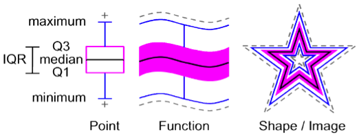
Similarly, analysis of airway geometry, which we represent as functions of cross-sectional area over the length of the airway, can be performed locally. However, to analyze airways in their entirety requires working with functions as data objects. This requires, for example, computing a median curve corresponding to the airway of an actual subject. In contrast, a point-wise median will result in a “median” curve which is not in the dataset and hence will “mix-and-match” data from different subjects.
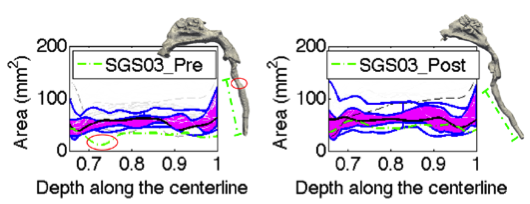
We have developed a general methodology based on functional boxplots for statistical atlas construction using curves as data objects [2] . We have applied this method to spatio-temporal atlas construction for pediatric airways and used the resulting atlas to to assess the severity of malformations in pediatric airways for subglottic stenosis.

Furthermore, our general image registration work has led to an improved method for the diagnosis of primary ciliary dyskinesia (PCD) [3], by exploiting the inherent symmetry of dynein arms to provide the pathologist cleaner images on which to base a diagnosis. We demonstrated that such an approach indeed results in diagnostic improvements.
References:
[1] J. Miedema, J.S. Marron, M. Niethammer, D. Borland, J. Woosley, J. Coposky, S. Wei, N.E. Thomas, “Image and Statistical Analysis of Melanocytic Histology,” Histopathology, 2012. [2] Y. Hong, B. Davis, J. Marron, R. Kwitt and M. Niethammer, “Weighted Functional Boxplot with Application to Statistical Atlas Construction,” accepted to MICCAI 2013. [3] W. K. Funkhouser III, M. Niethammer, J. L. Carson, K. A Burns, M. R. Knowles, M. W. Leigh, M. A. Zariwala and W. K. Funkhouser Jr., “A new tool improves diagnostic test performance for transmission EM evaluation of axonemal dynein arms.,” Ultrastructural Pathology, 2013, accepted for publication.