Fungipods

March 2, 2011

Aaron Neumann and his colleagues from Cell and Developmental Biology found novel dorsal pseudopodial protrusions, the “fungipods”, formed by dendritic cells (red objects in the upper image)  after contact with yeast cells (i.e. green blobs in the upper image).

Fungipods have a convoluted cell-proximal end and a smooth distal end. They persist for hours, and exhibit noticeable growth at the contact. Aaron Neumann et al. think that fungipods may promote yeast particle phagocytosis (i.e. process of surrounding and consuming solid particles) by dendritic cells.

Virtual Lung Video: Mucus River, Scene 2

September 19, 2009

Clearance2.mov

The second in a series of animated videos depicting the inner workings of the human lung on a microscopic scale. “Scene 2: Clearance: A Journey” asks questions about how clearance can possibly work when the volume through which the mucus flows decreases as it moves up from the depths of the lung to the throat.

This video can also be seen on YouTube.

Mitotic Spindle 3D Model Zoom

August 12, 2009
3D Spindle Model Close-up

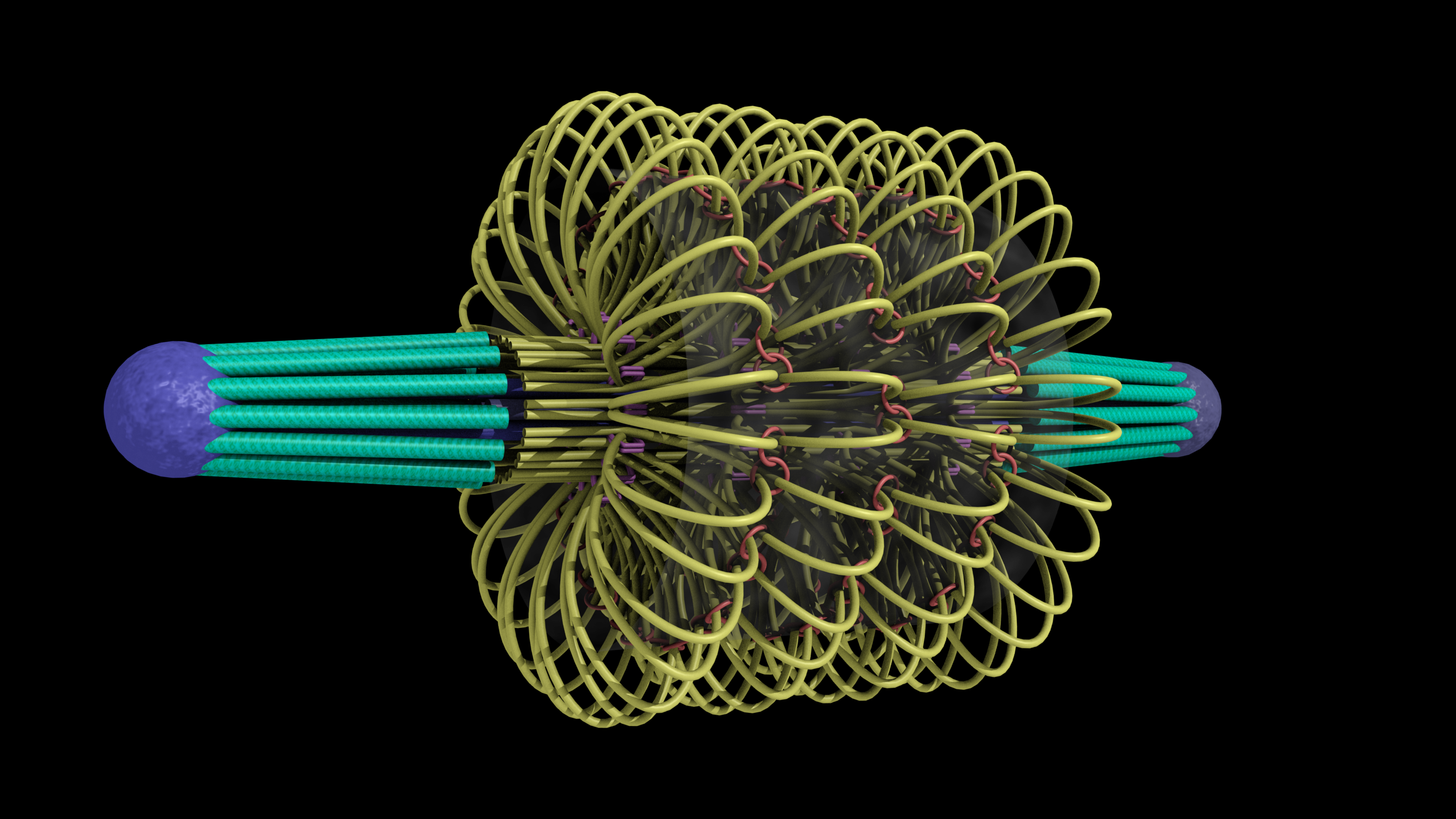
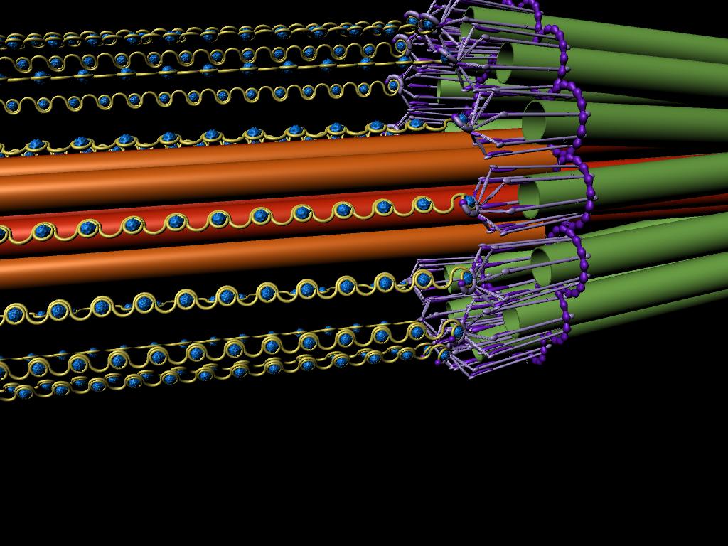
This is an image of Kendall McKenzie’s 3D model of the mitotic spindle made in Maya rendered from a zoomed-in perspective.

Mitotic Spindle Cross Section

July 23, 2009

This drawing shows a cross section of the mitotic spindle, with a diameter of 250 nm.  The 16 kinetochore microtubules are green, the 8 interpolar microtubules are orange, and the strands of DNA are represented by purple squiggly lines

Close-up Drawing of DNA Structure

July 23, 2009
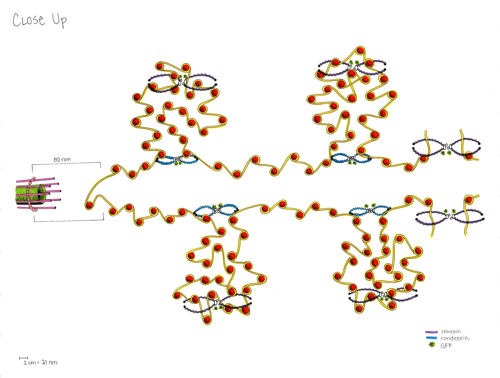
This drawing shows the form DNA takes when it is between the microtubule (green) end and a gap from which a loop extends. The microtubule is attached to the DNA by the kinetochore structure (pink), which is 80 nm long. The kinetochore attaches to the DNA (yellow) at a hook-like space near the cse4 nucleosome (shown at the end). There are 55 histones (orange) on each of the "top" and "bottom" of the structure. Roughly 75% of the length of the DNA strand is tangled in "loop" structures, and the other 25% is between loops in a straighter line. Condensin (blue) binds to the DNA at the bottoms of the loops. Condensin (purple) wraps around and encloses the DNA within the loops and where there is a space on the far right. The small green asterisks show where the GFPs are placed on cohesin and condensin in the lab.
This drawing shows the form DNA takes when it is between the microtubule (green) end and a gap from which a loop extends. The microtubule is attached to the DNA by the kinetochore structure (pink), which is 80 nm long. The kinetochore attaches to the DNA (yellow) at a hook-like space near the cse4 nucleosome (shown at the end). There are 55 histones (orange) on each of the “top” and “bottom” of the structure. Roughly 75% of the length of the DNA strand is tangled in “loop” structures, and the other 25% is between loops in a straighter line. Condensin (blue) binds to the DNA at the bottoms of the loops. Condensin (purple) wraps around and encloses the DNA within the loops and where there is a space on the far right. The small green asterisks show where the GFPs are placed on cohesin and condensin in the lab.

Mitotic Spindle

July 23, 2009
This drawing shows the spindle from end to end.  It is roughly 1.5 micons in length, with the kinetochore (green) microtubules making up 350 nm on each end, and the space between them being roughly 800 nm.  The interpolar microtubules are drawn in two shades different shades of purple (dark purple ones extend from the left end of the spindle and pink-purple ones extend from the right end of the spindle).  The microtubules are drawn straight and rigid, with an overall spindle diameter of 250 nm.   Also, the DNA strands (yellow) extend even more outwardly than the microtubules.  The two outermost strands of DNA (top and bottom of the spindle) have a few loops of DNA drawn in to show that the diameter of the spindle reaches 400 nm when the loops are included.  Each of the other strands has a small break in it that represents a place where extra loops of DNA are attached.  They are not drawn because they would make the details of the spindle hard to see. The bottom right corner shows the layout and packing of the microtubules at the spindle pole, not in a "convergence point" formation, but in a more tightly packed formation that has a diameter of 150 nm.
This drawing shows the spindle from end to end. It is roughly 1.5 micons in length, with the kinetochore (green) microtubules making up 350 nm on each end, and the space between them being roughly 800 nm. The interpolar microtubules are drawn in two shades different shades of purple (dark purple ones extend from the left end of the spindle and pink-purple ones extend from the right end of the spindle). The microtubules are drawn straight and rigid, with an overall spindle diameter of 250 nm. Also, the DNA strands (yellow) extend even more outwardly than the microtubules. The two outermost strands of DNA (top and bottom of the spindle) have a few loops of DNA drawn in to show that the diameter of the spindle reaches 400 nm when the loops are included. Each of the other strands has a small break in it that represents a place where extra loops of DNA are attached. They are not drawn because they would make the details of the spindle hard to see. The bottom right corner shows the layout and packing of the microtubules at the spindle pole, not in a “convergence point” formation, but in a more tightly packed formation that has a diameter of 150 nm.

Chromatin in the Nucleus

July 23, 2009

This drawing shows the packing of the chromatin (orange) in the nucelus.  The microtubules (green) extend out both sides of the nuclear membrane, and the chroma

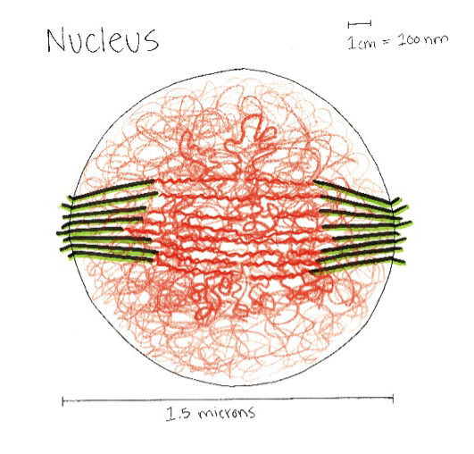
This drawing shows the packing of the chromatin (orange) in the nucelus.  The microtubules (green) extend out both sides of the nuclear membrane, and the chromatin is bunched/tangled up inside the nucleus.  The round nucleus is about 1.5 microns in diameter.
This drawing shows the packing of the chromatin (orange) in the nucelus. The microtubules (green) extend out both sides of the nuclear membrane, and the chromatin is bunched/tangled up inside the nucleus. The round nucleus is about 1.5 microns in diameter.

tin is bunched/tangled up inside the nucleus.  The round nucleus is about 1.5 microns in diameter.

FluoroSim: Interaction with specimen models

July 14, 2009

Simulated image generation in FluoroSim is fast enough to enable interaction with specimen models while watching real-time updates of the expected fluorescence image. This video shows some of FluoroSim’s main capabilities.

Click here for youtube video.

Mucus flow visualization

July 10, 2009
Cilia-driven mucus flow visualization by David Borland

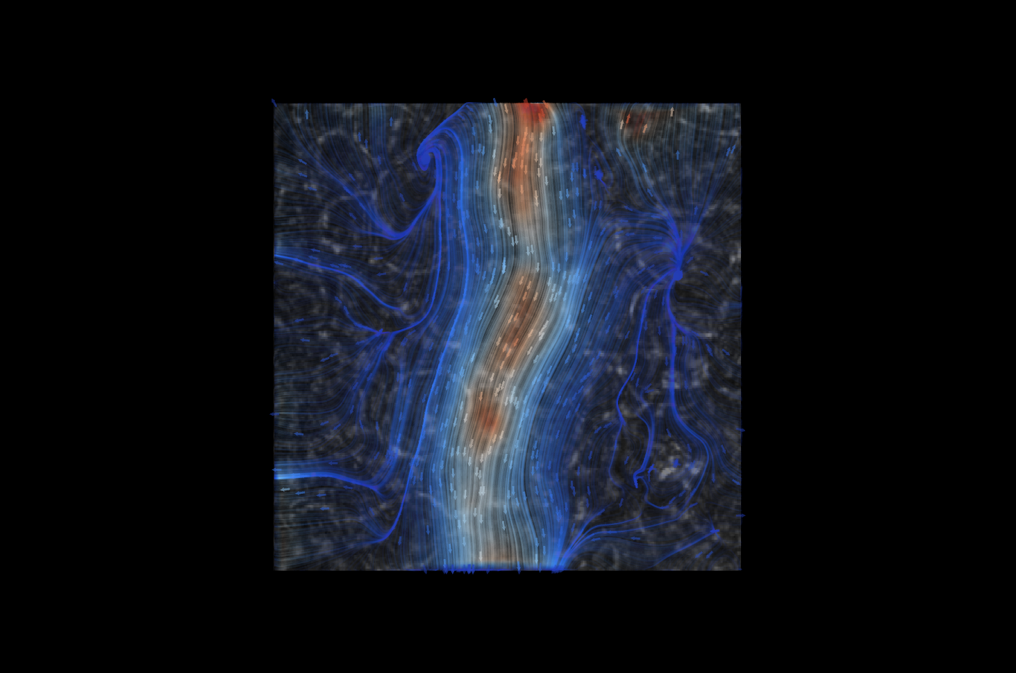
David Borland developed a flow visualization technique and used the output of Brian Eastwood’s ImageTracker program to construct this visualization of cilia-driven mucus flow on a human lung cell culture video from David Hill.  He overlaid the flow on the cell background determined by ImageTracker.

Flow speed is encoded both by color (blue lowest, through gray to red).  Flow direction is along the lines and in the direction pointed to by the arrows.

Virtual Lung Video: Mucus River, Scene 1

July 9, 2009

virtual-lung-final-video32

The first in a series of animated videos depicting the inner workings of the human lung on a microscopic scale. “Scene 1: Into the Mucus River” explains how air gets into healthy lungs, how the body defends itself against harmful airborne pathogens, and how mucus and cilia interact with the air particles.

This video can also be seen on YouTube.