ImageTracker Tutorial: Stationary Component Removal
This tutorial demonstrates how to remove stationary components from a bright field image sequence using ImageTracker. This method may be useful for digitally removing stationary specimen layers (e.g. cells grown on a substrate), removing debris on image sensors, and correcting for non-uniform intensity illumination (flat fielding).
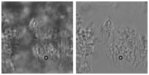
Data Set and Source
This data set is from Dr. David Hill at Rich Superfine’s laboratory at the University of North Carolina. In this video, captured at 120 frames per second, a bead is attached to beating cilia on epithelial lung cells. The stationary cell structure visible in the background confounds some of the motion present. Also, an artifact on the image sensor is visible in the middle of the image. In this tutorial, we will remove the effects of these stationary elements from the video.
Recommended Analysis Procedure
Select Process->Remove Occlusions from the ImageTracker menu. The default parameters work well for this example; the Maximum Transmission parameter adjusts the brightness of the output video. Ensure that you have write access to the output directory, especially if you loaded the data from a DVD. A progress bar monitors the process after pressing the Run button; when the process has finished, the resulting image sequence will automatically load in ImageTracker.
Note that a file called “transmission.mha” is saved to the output directory. This file contains a transmission map over the image plane for the stationary components in the image sequence. This file can be used in a Flatfield filter for any images taken using the same imaging conditions. It may be useful, for example, to compute the transmission map using a small set of images and apply that transmission map to a larger sequence.