Appearance Normalization for Histology Slides
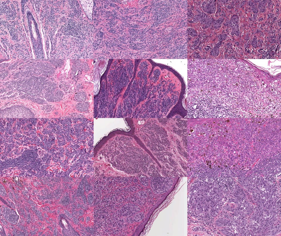
In the context of skin cancer we have developed an automatic method to normalize the appearance of H&E-stained histology slides [1,2]. This is a critical preprocessing step for subsequent quantitative image analysis to minimize the influence of differences in staining (due to different staining protocols, different ages of slides, etc.) on an analysis result. The method both estimates the stain vectors for hematoxylin and the eosin stains as well as overall stain intensities and transforms them into a pre-specified space for normalization. Prior information on the stains can be incorporates for increased robustness of the estimator. The method is fully automatic and we have used it successfully on hundreds of histology slides.

References:
[1] M. Macenko, M. Niethammer, J. S. Marron, D. Borland, J. T. Woosley, X. Guan, C. Schmitt, and N. E. Thomas, “A method for normalizing histology slides for qunatitiative analysis,” ISBI, 2009, pp. 1107-1110. [2] M. Niethammer, D. Borland, S. J. Marron, J. Woosley, and N. E. Thomas, “Appearance normalization of histology slides,” MICCAI Machine Learning in Medical Imaging Workshop, 2010, pp. 58-66.