Fungipod

April 1, 2012

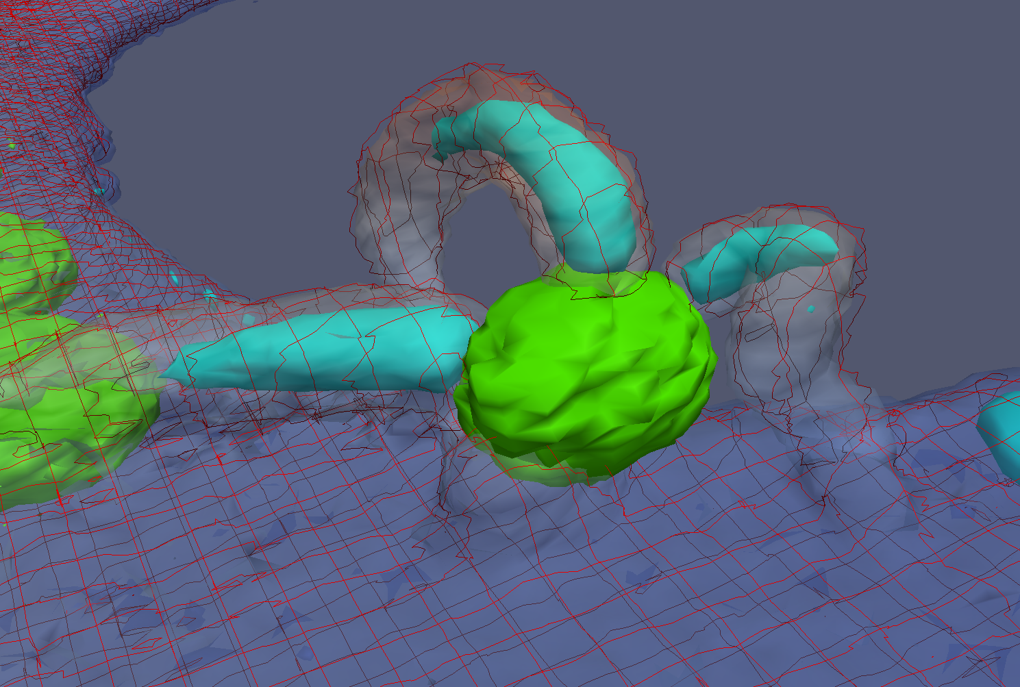
Actin-rich fungipods attack a yeast attached to a cell membrane

Collaborator Aaron Neumann from the University of New Mexico is studying fungipods from human dendritic cells that attach to yeast.  The image above shows a combination of three different fluorophores that together show the behavior.  A green yeast is sitting on top of the cell membrane (transparent red) with three fungipods attached to it.  The fungipods are very dense in actin (blue).
FluoroSim: Interaction with specimen models

July 14, 2009

Simulated image generation in FluoroSim is fast enough to enable interaction with specimen models while watching real-time updates of the expected fluorescence image. This video shows some of FluoroSim’s main capabilities.

Click here for youtube video.

Mucus flow visualization

July 10, 2009
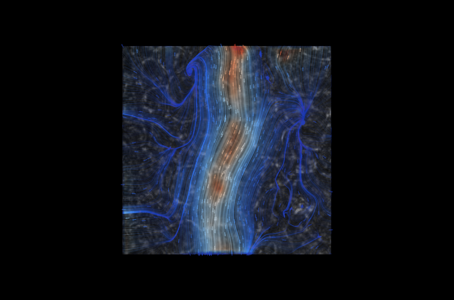
Cilia-driven mucus flow visualization by David Borland

David Borland developed a flow visualization technique and used the output of Brian Eastwood’s ImageTracker program to construct this visualization of cilia-driven mucus flow on a human lung cell culture video from David Hill.  He overlaid the flow on the cell background determined by ImageTracker.

Flow speed is encoded both by color (blue lowest, through gray to red).  Flow direction is along the lines and in the direction pointed to by the arrows.

ImageSurfer clarifies cilia locations

July 2, 2009
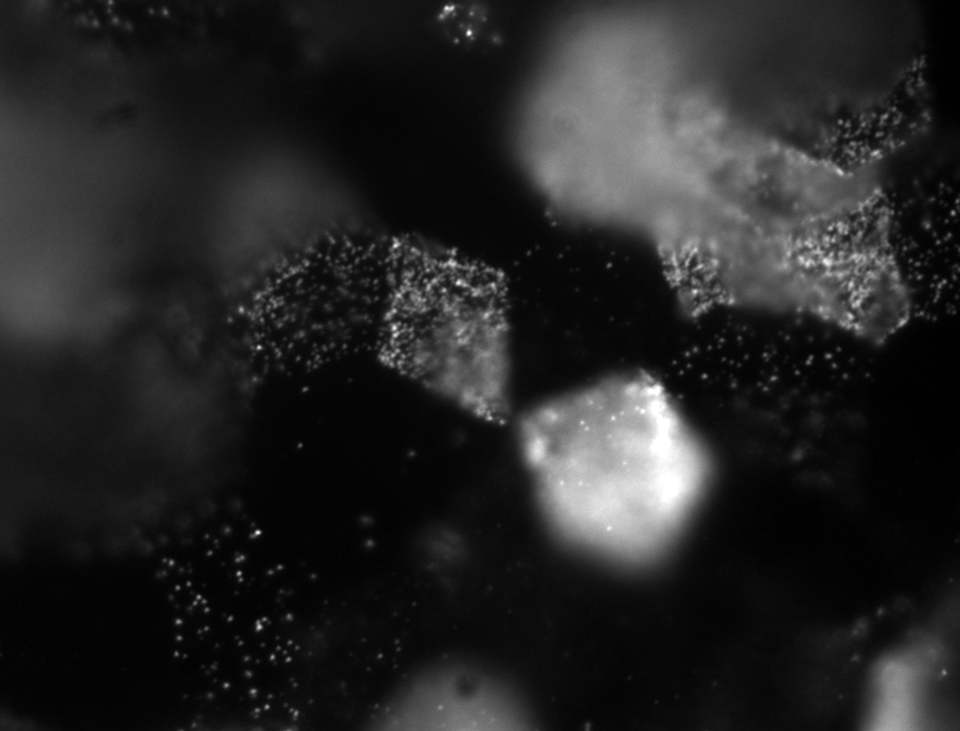
Slice through epithelial cell culture.

The image above shows one frame from a stack of images taken of a human lung epithelial cell culture.  The well-separated spots were thought to be cross sections through cilia sticking out the tops of cells.  There are some bright spots on the labeled cells and others (near the left of the image) that were thought to be above cells.

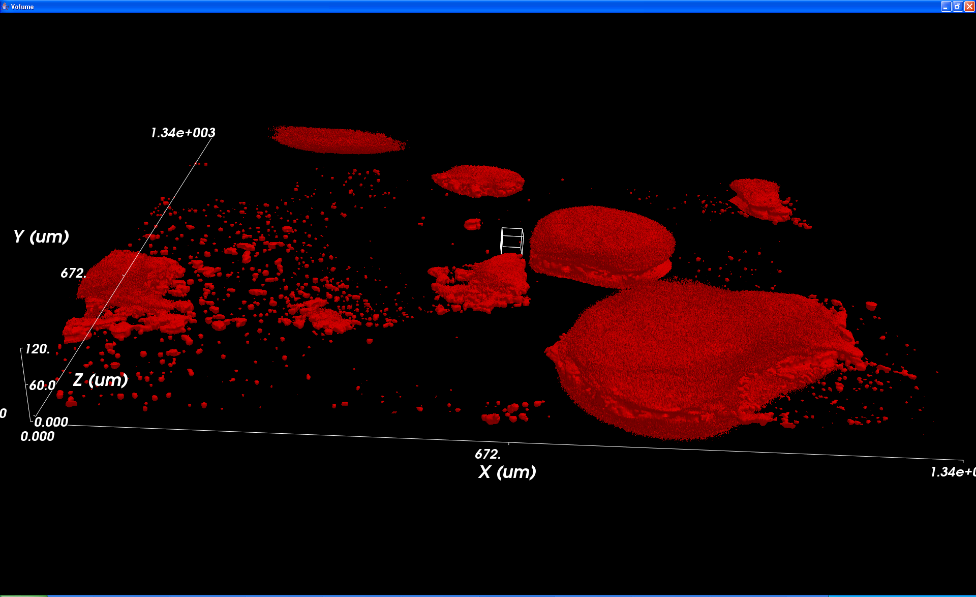
The ImageSurfer image shown below  revealed that they were in fact isolated blobs of dye that were outside of the cells.

ImageSurfer 3D view of epithelial cells